
InsightsHealthcare
AI-Powered Skin Cancer Detection Apps: Development Guide 2026
Learn how to build a clinically validated AI skin cancer detection app in 2026—covering FDA compliance, tech stack, costs, and real implementation insights.
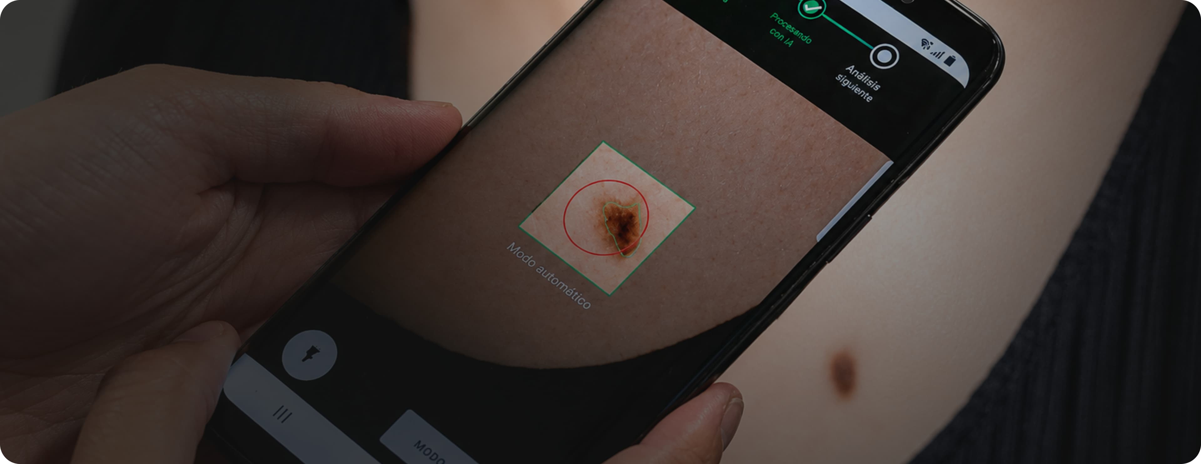
Melanoma caught at stage one has a five-year survival rate above 98%. Melanoma caught at stage four has a five-year survival rate below 25%. The difference between those two outcomes is almost entirely about when the cancer is detected, and detection almost always starts with someone noticing a suspicious lesion and deciding whether to seek evaluation.
Most people do not seek evaluation when they should. The barrier is not ignorance of the risk. It is access to a dermatologist, to an appointment, to a diagnosis. In the United States, the median wait time to see a dermatologist exceeds 30 days in most markets. In rural areas, it can exceed six months. By the time a patient gets to an appointment, a lesion that was suspicious three months ago may have changed significantly.
An AI skin cancer detection app changes the triage decision. It gives anyone with a smartphone the ability to photograph a suspicious lesion, receive an AI-powered assessment of whether it warrants urgent dermatological evaluation, and, in systems with clinical integration, connect directly to a teledermatology platform for professional review.
In 2026, AI skin cancer detection is one of the most clinically validated and most actively deployed categories of healthcare AI. The technology has been validated in peer-reviewed clinical studies, several tools have received FDA clearance, and both direct-to-consumer and clinical-grade platforms are in active use across the United States and internationally.
This guide covers everything healthcare startups and digital health companies need to know about building an AI skin cancer detection app from the clinical validation requirements to the technical architecture, the FDA regulatory pathway, the HIPAA compliance obligations, and the step-by-step development process.
Key Takeaways
An AI skin cancer detection app uses computer vision and deep learning to analyze images of skin lesions and assess their likelihood of malignancy, helping patients triage whether a lesion warrants urgent dermatological evaluation
Clinical validation against a dermatologist reference standard is a non-negotiable requirement; sensitivity and specificity must be validated on demographically representative datasets before any clinical deployment
FDA regulatory classification is critical to address before development begins. AI skin cancer detection tools that make diagnostic or triage claims are regulated as Software as a Medical Device and may require 510(k) clearance or De Novo classification
HIPAA compliance is required for any app that connects to healthcare providers or handles identifiable patient health information
Demographic representation in training data is a clinical equity requirement; AI models trained on non-representative datasets perform less accurately for underrepresented skin tones and demographics
The most successful AI skin cancer detection platforms in 2026 combine AI triage with teledermatology integration. The AI determines urgency; the dermatologist makes the diagnosis
Total development cost ranges from $80,000 for a basic consumer screening app to $500,000 or more for a clinically validated, FDA-cleared platform
What Is an AI Skin Cancer Detection App?
An AI skin cancer detection app is a digital health tool that uses computer vision, specifically deep learning models trained on large datasets of labeled dermoscopic and clinical photographs to analyze images of skin lesions and provide an assessment of their likelihood of malignancy.
The app captures an image of a skin lesion through the device camera, either a standard smartphone camera for clinical photographs or a dermatoscope attachment for dermoscopic images, and applies AI models to assess visual features associated with different types of skin lesions. Outputs typically include a risk classification such as the likelihood of melanoma versus a benign lesion, a recommendation for the appropriate clinical follow-up based on the risk assessment, and visual highlights of the specific lesion features that influenced the AI assessment.
The critical clinical and regulatory distinction is between an app that provides triage guidance recommending whether a lesion warrants urgent, routine, or no professional evaluation and one that provides a diagnosis. AI skin cancer detection apps are screening and triage tools. They do not replace dermatological diagnosis. They make the triage decision faster, more accessible, and more accurate than the unaided patient judgment that currently determines whether most people seek evaluation.
This distinction has direct regulatory implications. Apps that make triage recommendations rather than diagnostic claims may have a different FDA regulatory pathway than apps that claim to diagnose specific conditions, though this determination requires regulatory counsel assessment for each specific intended use.
Why Is AI Skin Cancer Detection One of the Most Validated Healthcare AI Applications?
Several factors have made AI skin cancer detection one of the most clinically robust categories of AI in medicine.
The data is available and well-labeled. Dermoscopic image archives from academic dermatology centers contain hundreds of thousands of labeled skin lesion images, including positive and negative cases for melanoma, basal cell carcinoma, squamous cell carcinoma, and benign conditions including seborrheic keratosis, dermatofibromas, and benign nevi. The ISIC Archive, the International Skin Imaging Collaboration, has been instrumental in building and sharing standardized training datasets.
The task is well-defined and measurable. Image classification for skin lesion malignancy is a well-scoped computer vision problem with clear ground truth; the pathological diagnosis from a biopsy specimen provides the definitive label for model training and validation. This clarity makes clinical validation straightforward compared to AI tasks with more ambiguous ground truth.
The clinical need is enormous. Skin cancer is the most common cancer in the United States. More than five million cases are diagnosed annually. Dermatologist access is severely constrained. The gap between the scale of the problem and the availability of specialist access is exactly the kind of problem that AI screening tools are designed to address.
Early studies demonstrated clinical validity. Multiple peer-reviewed studies published between 2017 and 2026 have demonstrated that AI skin lesion classification achieves diagnostic accuracy comparable to dermatologist-level performance for specific lesion types, most notably the 2017 Esteva et al. Nature paper that demonstrated CNN-based skin lesion classification at a level comparable to board-certified dermatologists.
What Are the Types of AI Skin Cancer Detection Apps?
1. Consumer Screening Apps
Direct-to-consumer apps that allow patients to photograph suspicious lesions and receive AI risk assessments without clinician involvement. These apps operate in a regulatory space that requires careful intended use definition; they can provide general skin health information but must be designed to avoid making diagnostic claims that would trigger FDA regulation as a medical device without clearance.
Consumer screening apps are most valuable as tools that lower the threshold for seeking professional evaluation, prompting users who might otherwise ignore a suspicious lesion to consult a dermatologist.
2. Clinical-Grade Screening Tools for Primary Care
AI skin lesion assessment tools designed for use by primary care physicians, nurse practitioners, and physician assistants who encounter skin concerns during routine care but do not have dermatology specialist training. These tools provide AI-assisted assessment that helps primary care providers make more informed triage decisions, referring urgently, referring routinely, or monitoring in-office rather than relying on unaided clinical judgment.
Clinical-grade tools designed for use by healthcare providers are more likely to be regulated as Software as a Medical Device than consumer apps because they are positioned as clinical decision support rather than general health information.
3. Teledermatology Integration Platforms
Apps that combine AI-powered lesion assessment with integration to a teledermatology platform enabling a patient who receives a concerning AI risk assessment to immediately connect with a dermatologist for professional review without leaving the app. This model AI triage plus teledermatology diagnosis represents the most complete clinical workflow for AI skin cancer detection.
4. Dermatologist Decision Support Tools
AI tools designed for use by dermatologists during clinical evaluation, augmenting the dermatologist's assessment with AI analysis of dermoscopic images. These tools are designed to improve diagnostic accuracy and consistency, not to extend access to non-specialists.
5. Population Screening Programs
AI skin cancer detection is deployed as part of organized population screening programs in pharmacies, employee health programs, community health events where standardized imaging is performed, and AI assessment is used to triage individuals for follow-up dermatological evaluation.
What Are the Clinical Validation Requirements?
Clinical validation is the requirement that most distinguishes AI skin cancer detection app development from general consumer health app development, and it is the requirement that most healthcare startups underestimate.
Validation Against a Dermatologist Reference Standard
For any AI skin cancer detection tool intended for clinical use, whether by patients for triage or by providers for decision support, validation against a dermatologist or pathological reference standard is required. This means demonstrating that the AI achieves acceptable sensitivity and specificity on an independent test set, one that was not used in training or hyperparameter optimization.
Sensitivity, the rate at which the AI correctly identifies malignant lesions, and specificity, the rate at which the AI correctly classifies benign lesions, are the primary validation metrics. For skin cancer screening specifically, sensitivity is typically the more critical metric; missing a malignant lesion is a worse outcome than a false positive that prompts an unnecessary dermatologist visit.
Demographic Representation in Training and Validation Datasets
AI skin cancer detection models trained primarily on images of patients with light skin tones perform significantly less accurately on patients with darker skin tones because the visual features of melanoma present differently across skin tone ranges. This is not a minor performance difference. It is a health equity concern that has been documented in published studies of AI dermatology tools.
Building a training dataset that adequately represents the demographic diversity of the intended patient population, including skin tones across the full Fitzpatrick scale, is a clinical equity requirement and an emerging regulatory expectation. Validation must include subgroup analysis by skin tone and demographic group to demonstrate that the model does not have clinically significant performance disparities across the patient population it will serve.
Validation for Each Lesion Type and Clinical Context
A model that achieves excellent sensitivity and specificity for melanoma on dermoscopic images may perform significantly less well on clinical photographs or on non-melanoma skin cancers or in specific body site locations. Validation must cover the specific lesion types, image types, and clinical contexts for which the app will be used.
What Are the Key Features of an AI Skin Cancer Detection App?
High-Quality Image Capture
The clinical accuracy of any AI skin cancer detection app depends on the quality of the input image. Image capture guidance instructions to the user on optimal lighting, distance, focus, and angle significantly reduce the proportion of images that are too poor quality for reliable AI analysis.
For consumer apps using standard smartphone cameras, image quality guidance is the most impactful design feature for improving AI accuracy in real-world conditions. For clinical apps using dermatoscope attachments, integration with the specific hardware is required.
AI Lesion Analysis Engine
The core AI capability is a deep learning model trained on labeled dermoscopic and clinical photographs that classifies the input image by lesion type and malignancy likelihood. The model architecture, training data, and validation results are the determinants of clinical accuracy.
Our AI and ML solutions team builds skin lesion classification models with the training data diversity, validation rigor, and explainability outputs, including GradCAM heatmaps showing the image regions driving the AI assessment that clinically responsible skin cancer detection requires.
Risk Classification and Clinical Recommendation
AI assessment output presented to the user in clinically meaningful terms: risk level, recommended action, and explanation of the basis for the recommendation. The language used to present risk classification must be carefully designed to be actionable without making claims that overstate the AI's diagnostic capability.
Lesion Tracking and Longitudinal Monitoring
For users monitoring lesions over time, the ability to photograph the same lesion at multiple time points and track changes in size, shape, color, and AI risk classification provides clinically meaningful longitudinal information that a single assessment cannot.
Dermoscopic studies have demonstrated that lesion change over time is a meaningful predictor of malignancy. AI tools that quantify and track change add clinical value beyond single-point assessment.
Teledermatology Integration
Integration with a teledermatology platform that enables users who receive concerning AI assessments to connect directly with a board-certified dermatologist for professional evaluation. This integration, combining AI triage with professional diagnosis, is what makes the AI skin cancer detection workflow clinically complete rather than stopping at triage.
Our API integration services team builds integrations with teledermatology platforms using the data formats and communication protocols required to transfer lesion images, AI assessment results, and patient information securely.
Explainability and Visual Highlighting
AI assessment output that shows the user or the clinician which visual features of the lesion are driving the AI's risk classification. GradCAM visualization overlaid on the lesion image shows which regions the model focused on. Feature importance scores indicate which dermoscopic features were most influential.
Explainability is both a clinical trust requirement for clinicians to understand the basis for AI recommendations to evaluate whether to act on them and a regulatory expectation for AI medical devices that influence clinical decisions.
HIPAA-Compliant Data Handling
For apps that connect users with healthcare providers or store identifiable patient health information alongside lesion images, HIPAA compliance is required. Lesion images combined with identifying information name, date of birth, contact information constitute protected health information.
Our HIPAA-compliant software development practice builds the compliance architecture encryption, access controls, audit logging, and BAA management appropriate for digital health apps that handle sensitive patient data.
Clinical Dashboard for Dermatology Practices
For AI skin cancer detection tools integrated into clinical workflows, a dermatologist-facing dashboard showing submitted cases, AI assessments, case priority by risk classification, and teledermatology consultation status provides the clinical team interface required for efficient case review.
Our healthcare UI/UX design team designs dermatologist dashboards tested with real dermatologists in realistic clinical review scenarios.
How to Build an AI Skin Cancer Detection App: Step by Step?
Step 1: Define the Intended Use and Target Population
Building an AI skin cancer detection app begins with a precisely defined intended use: who will use the app, in what clinical context, to make what decision. The intended use statement shapes the FDA regulatory pathway, the clinical validation requirements, the training data requirements, and the design of the risk communication to users.
Be specific. A consumer app that helps patients decide whether to seek dermatological evaluation has a different intended use than a clinical tool that helps primary care providers triage referrals, which has a different intended use than a decision support tool for dermatologists reviewing dermoscopic images.
Step 2: Determine the FDA Regulatory Pathway
Before any development begins, determine whether your intended use requires FDA clearance and what regulatory pathway applies.
AI skin cancer detection tools that make diagnostic claims identifying a lesion as malignant are regulated as Software as a Medical Device and require FDA clearance. Tools that provide general skin health information without diagnostic claims may fall outside FDA jurisdiction, but this determination requires regulatory counsel assessment based on the specific intended use.
Several FDA-cleared AI skin cancer detection tools exist as potential 510(k) predicates, including MelaFind, SciBase Nevisense, and DermEngine, which may support a 510(k) substantial equivalence argument for tools with similar intended uses.
Our MVP and product strategy process addresses FDA regulatory classification as a core component of the discovery phase because this determination affects every subsequent development decision.
Step 3: Assemble the Training Dataset
The clinical performance of an AI skin cancer detection model is entirely determined by the training dataset. Requirements include sufficient volume, typically tens of thousands of labeled images for robust model training, demographic diversity across skin tones and patient demographics, pathological confirmation of ground truth labels where possible, and coverage of the lesion types and image types for which the app will be used.
Public datasets, the ISIC Archive, HAM10000, and the MSKCC dataset provide important training data foundations. Proprietary clinical datasets from dermatology practices provide the demographic diversity and lesion type coverage that public datasets may lack for specific patient populations.
Step 4: Develop and Validate the AI Model
Build and train the deep learning model for skin lesion classification, typically using convolutional neural networks or transformer-based vision architectures. Transfer learning from models pre-trained on large image datasets EfficientNet, ResNet, Vision Transformer is standard practice, with fine-tuning on the assembled skin lesion dataset.
Implement explainability outputs, such as GradCAM or similar gradient-based visualization, as part of the model architecture, not as a post-hoc addition.
Validate model performance against an independent test set drawn from the same patient population, calculating sensitivity, specificity, AUC-ROC, positive predictive value, and negative predictive value. Conduct subgroup analysis by skin tone, lesion type, body site, and image type.
Step 5: Design the Risk Communication Framework
Design the language, visual presentation, and recommended action guidance associated with each risk classification output. This is a clinical communication design task that requires both clinical input to ensure the recommendations are clinically appropriate and patient communication expertise to ensure the language is comprehensible and actionable for users without medical training.
Risk communication must be designed to avoid overclaiming, presenting AI triage as diagnosis while being clear enough to prompt appropriate action for concerning assessments.
Step 6: Build the Image Capture and Quality Assessment Layer
Build the image capture interface with guidance overlays that help users photograph lesions at optimal quality distance indicators, lighting guidance, focus confirmation, and image quality scoring that flags poor-quality images for retake before submitting to the AI model.
Implement automatic image quality assessment using a quality classifier trained to identify images that are too blurry, too dark, too far away, or otherwise unsuitable for reliable AI analysis and reject or flag poor-quality images before AI assessment.
Step 7: Build Teledermatology Integration
Build integration with the teledermatology platform securely transferring lesion images, AI assessment results, and relevant patient information to the dermatology review platform, and receiving consultation results back into the app for display to the user.
Step 8: Implement HIPAA Compliance Architecture
For apps that handle identifiable patient health information, which includes lesion images combined with patient identifiers, any app connecting to clinical services should implement full HIPAA compliance architecture before any patient data is processed.
Step 9: Design the Patient Interface
Design the patient-facing app interface image capture flow, AI assessment presentation, risk communication, and teledermatology connection for the full range of the intended user population. Skin cancer affects patients across all age groups. The interface must be accessible and usable for users who are not digitally confident as well as those who are.
Step 10: Conduct FDA Pre-Submission and Regulatory Preparation
Before submission, engage with the FDA through the pre-submission process by submitting a pre-sub request that describes the intended use, the proposed regulatory pathway, and the clinical validation study design. FDA feedback on pre-sub requests guides the clinical validation approach and submission preparation.
Step 11: Pilot and Post-Market Surveillance
Deploy in a structured clinical pilot with defined performance metrics: sensitivity and specificity in real-world use, image quality rates, user engagement rates, referral rates, and dermatologist concordance with AI risk classifications.
Build post-market surveillance infrastructure from the start, monitoring model performance in production for drift and demographic performance disparities, and maintaining the performance documentation required for FDA post-market requirements.
Our DevOps and cloud solutions team builds the deployment infrastructure, model monitoring, and post-market performance tracking that regulatory responsibility for an AI medical device requires.
What Technology Powers an AI Skin Cancer Detection App?
Computer Vision and Deep Learning
PyTorch is the standard deep learning framework for skin lesion classification model development. EfficientNet, ResNet, and Vision Transformer architectures are the primary model families used in current state-of-the-art skin lesion classification systems, each with different trade-offs between accuracy, computational efficiency, and explainability.
For multi-class classification across skin cancer types melanoma, basal cell carcinoma, squamous cell carcinoma, actinic keratosis, and benign conditions multi-label classification architectures trained on large dermoscopic datasets produce the most clinically useful outputs.
MONAI Medical Open Network for AI provides pre-built components for medical image AI development that are relevant for clinical-grade skin lesion classification. GradCAM and Score-CAM provide gradient-based visualization methods for the explainability outputs required for clinical trust.
Image Processing
OpenCV provides the image processing utilities for quality assessment, standardization, and preprocessing. For dermoscope-captured images, color calibration and hair artifact removal preprocessing improve model accuracy in clinical settings.
Automated image quality classifiers: lightweight CNNs trained to detect blurry, dark, poorly framed, or otherwise unsuitable images provide the real-time quality feedback that guides users to capture images of sufficient quality for reliable AI analysis.
Mobile Application
React Native for cross-platform iOS and Android deployment is the standard choice for most AI skin cancer detection consumer apps, enabling single codebase development for both platforms while providing access to native camera APIs required for image capture.
For apps requiring dermoscope hardware integration, platform-specific native development in Swift for iOS and Kotlin for Android may be required to access the hardware APIs of specific dermatoscope attachment manufacturers.
Backend and Model Serving
Python with FastAPI handles the primary API layer. TensorFlow Serving or TorchServe provides model inference serving at the latency required for real-time image analysis. AWS Lambda or containerized API services handle the API layer for consumer-scale image submission volumes.
For apps that must provide AI assessment results within seconds of image submission, which is the user experience expectation for consumer apps, model serving infrastructure must be optimized for inference latency, not just throughput.
Cloud Infrastructure
AWS with HIPAA Business Associate Agreement for apps that handle protected health information. Amazon S3 with server-side encryption for secure lesion image storage. Amazon SageMaker for model training and deployment. AWS CloudTrail for HIPAA audit logging.
For apps that do not connect to clinical services and do not handle identifiable health information alongside images, on-device model inference using Core ML on iOS or TensorFlow Lite on Android eliminates the need to transmit lesion images to cloud servers, which may be preferable for consumer privacy and latency reasons.
Teledermatology Integration
HL7 FHIR R4 for clinical data exchange where teledermatology platforms support FHIR. Proprietary platform APIs for teledermatology platforms that use custom data formats. DICOM for dermatoscope image transfer in clinical environments that use DICOM-compatible imaging infrastructure.
What Are the FDA Regulatory Requirements for AI Skin Cancer Detection Apps?
Software as a Medical Device Classification
The FDA's Software as a Medical Device framework classifies software that meets the definition of medical device software intended to diagnose, cure, mitigate, treat, or prevent disease. AI skin cancer detection tools that make or support diagnostic claims about skin lesion malignancy are SaMD.
The specific regulatory pathway depends on the risk classification of the device and the existence of a substantially equivalent cleared predicate.
510(k) Clearance
The 510(k) pathway requires demonstrating that the device is substantially equivalent to a legally marketed predicate device in intended use and technological characteristics. Several AI dermatology tools have received 510(k) clearance, providing potential predicates for new submissions with similar intended uses.
De Novo Classification
For AI skin cancer detection tools with novel intended uses that do not have a substantially equivalent predicate, the De Novo pathway creates a new regulatory classification. De Novo submissions require demonstrating safety and effectiveness through clinical evidence rather than substantial equivalence.
Clinical Evidence Requirements
FDA submissions for AI skin cancer detection devices require clinical evidence demonstrating the device's sensitivity and specificity in the intended use population, typically from a prospective or retrospective reader study comparing the device to a dermatologist or pathological reference standard.
The clinical study design, number of cases, case mix, reference standard, reader qualification, and statistical analysis plan should be discussed with the FDA through the pre-submission process before the study is conducted.
Algorithm Transparency and Documentation
FDA submissions for AI medical devices require documentation of the model architecture, training data, training process, and validation methodology sufficient for FDA reviewers to evaluate the scientific validity of the AI system. This documentation must be prepared as part of the submission package and maintained as a controlled record.
What Are the HIPAA Compliance Requirements?
HIPAA applies to AI skin cancer detection apps that handle protected health information, lesion images combined with identifiable patient information, clinical data linked to identifiable users, and communications with healthcare providers about specific patients.
Consumer apps that analyze lesion images without associating them with identifiable patient information operating in a mode where the app cannot link the image to a specific individual may fall outside HIPAA's scope as a general wellness tool. This determination depends on the specific data architecture of the app and should be reviewed by legal counsel.
Apps that connect users with dermatologists, store lesion images with patient identifiers, or communicate with healthcare providers about specific patients are handling protected health information and must implement full HIPAA technical safeguards: encryption at rest and in transit, access controls, audit logging, and Business Associate Agreements with all third-party services.
AI Skin Cancer Detection App Development Checklist
Clinical and Regulatory Foundation
Intended use defined with precision: patient population, clinical context, decision supported
FDA regulatory classification determined before development begins
Pre-submission meeting with FDA scheduled for devices requiring clearance
Clinical validation study design reviewed by regulatory counsel
Training Data and Model Development
Training dataset assembled with demographic diversity across skin tones
Pathological or dermatologist ground truth labels confirmed
Model trained and validated on held-out independent test set
Subgroup analysis conducted by skin tone, lesion type, and body site
GradCAM or equivalent explainability outputs implemented
Image quality classifier implemented for real-time quality feedback
Clinical Validation
Sensitivity and specificity validated against dermatologist reference standard
Performance validated across demographic subgroups
Real-world image quality impact on performance assessed
Risk communication language clinically reviewed
Compliance
FDA regulatory submission prepared if required
HIPAA compliance architecture implemented for apps handling PHI
BAAs in place with all third-party services
Post-market surveillance infrastructure built
Product and Integration
Image capture guidance tested with representative user population
Teledermatology integration built and validated
Patient interface tested with users across age and digital confidence range
Clinical dashboard built and tested with dermatologists
What Are the Common Mistakes to Avoid?
1. Treating Clinical Validation as a Post-Development Task
Clinical validation for a skin cancer detection app is not something that happens after the product is built; it shapes what is built. The training dataset requirements, the model architecture, the intended use definition, and the FDA regulatory pathway all flow from the clinical validation requirements. Teams that treat validation as a post-development step consistently face expensive rebuilds when validation reveals that the product they built does not meet clinical standards.
2. Building on Non-Representative Training Data
AI skin cancer detection models trained primarily on images from patients with light skin tones perform significantly less accurately on patients with darker skin tones. This is not a theoretical concern; it has been documented in published clinical studies. Building on non-representative training data produces a product with significant health equity problems and potential regulatory risk.
3. Overclaiming in User-Facing Risk Communication
Risk communication language that implies the AI is providing a diagnosis "your lesion is likely melanoma" rather than a triage assessment creates both clinical harm potential and regulatory risk. Risk communication must be precisely designed to be actionable for users while accurately representing what the AI assessment can and cannot conclude.
4. Skipping FDA Regulatory Determination
The boundary between a general wellness app and a Software as a Medical Device is not always obvious for AI skin cancer detection tools. Assuming that a consumer skin screening app falls outside FDA jurisdiction without regulatory counsel assessment is a common and expensive mistake. The cost of regulatory determination before development is trivial compared to the cost of discovering a regulatory requirement after the product is built.
5. No Image Quality Filtering
AI skin cancer detection models produce unreliable outputs on low-quality input images, but they produce some output, not an error message. Users who submit low-quality images and receive AI assessments based on those images may act on assessments that do not reflect the actual lesion. Image quality filtering that identifies and rejects unsuitable images before AI analysis is a clinical safety requirement, not a polish feature.
How Codieshub Builds AI Skin Cancer Detection Apps?
At Codieshub, we build AI skin cancer detection apps for healthcare startups and digital health companies that need clinical-grade computer vision platforms with the demographic representation, clinical validation rigor, regulatory compliance, and explainability that clinically responsible skin cancer detection requires.
Every engagement begins with our MVP and product strategy process, which addresses intended use definition, FDA regulatory classification, training data strategy, clinical validation design, and HIPAA compliance architecture before production code is written. For AI skin cancer detection specifically, the decisions made before development begins determine whether the resulting product can be legally deployed and clinically trusted.
Our AI and ML solutions team builds skin lesion classification models with demographically representative training data, comprehensive clinical validation including demographic subgroup analysis, GradCAM explainability outputs, and image quality classification that ensures AI assessment is only applied to images of sufficient quality. Our healthcare UI/UX design team designs patient-facing image capture flows and risk communication interfaces tested with users across the full range of the intended patient population.
Our API integration services team builds teledermatology platform integrations that complete the clinical workflow from AI triage to professional diagnosis. Our HIPAA-compliant software development practice ensures full compliance for apps that handle protected health information. And our DevOps and cloud solutions team builds the deployment infrastructure, model monitoring, and post-market performance tracking that FDA post-market surveillance requirements demand.
Conclusion
AI skin cancer detection represents one of the most clinically validated and most impactful applications of computer vision in healthcare. The technology works. The clinical evidence supports it. And the gap it addresses between the scale of skin cancer incidence and the accessibility of dermatological expertise is real, consequential, and growing.
Building an AI skin cancer detection app that is clinically responsible with demographically representative training data, rigorous clinical validation, appropriate FDA regulatory compliance, and risk communication designed to inform rather than mislead is significantly more demanding than building a general consumer health app. But it is exactly this clinical rigor that produces a product that actually improves patient outcomes rather than one that generates downloads without clinical impact.
At Codieshub, we build AI skin cancer detection apps for healthcare startups and digital health companies that are committed to clinical responsibility with the computer vision expertise, clinical validation rigor, FDA regulatory navigation, and HIPAA compliance architecture that building in this domain demands.
Ready to build an AI skin cancer detection app that is both clinically valid and regulatory ready?
Schedule a Discovery Call to tell us about your intended use and target population, and we will send you a tailored development and regulatory game plan within 48 hours.
Frequently Asked Questions
1. What Is an AI Skin Cancer Detection App?
An AI skin cancer detection app uses deep learning and computer vision to analyze photographs of skin lesions and assess their potential risk. It helps users decide whether a lesion may require professional evaluation. The app supports screening and triage but does not replace a dermatologist's diagnosis.
2. How Accurate Are AI Skin Cancer Detection Apps?
AI skin cancer detection models can achieve dermatologist-level sensitivity and specificity for certain lesion types when validated on high-quality datasets. However, real-world accuracy can vary because of image quality, photography conditions, and dataset diversity. Models trained on representative data generally provide more reliable results across different skin tones.
3. Do AI Skin Cancer Detection Apps Need FDA Clearance?
FDA clearance depends on the app's intended use and claims. Apps making diagnostic or specific triage claims may be regulated as Software as a Medical Device and could require 510(k) clearance or De Novo classification. Regulatory requirements should be assessed before development with qualified regulatory counsel.
4. Does an AI Skin Cancer Detection App Need to Be HIPAA Compliant?
HIPAA applies when an app handles protected health information, such as identifiable patient details combined with lesion images or clinical communications. Apps connecting users with dermatologists or healthcare providers typically require appropriate safeguards, including encryption, access controls, audit logging, and Business Associate Agreements with relevant service providers.
5. What Training Data Does an AI Skin Cancer Detection Model Need?
Effective AI skin cancer models require large collections of labeled clinical or dermoscopic images with reliable dermatologist or pathological ground truth. Training data should represent different skin tones, demographics, lesion types, and clinical contexts. Public datasets such as the ISIC Archive can provide a foundation for model development.
6. What Is the Most Important Clinical Validation Requirement?
One of the most important requirements is validating model performance across demographic subgroups. Sensitivity and specificity should be assessed across different skin tones, patient demographics, lesion types, and clinical contexts. This helps identify performance disparities and ensures the model provides reliable results for the intended patient population.
7. How Long Does It Take to Build an AI Skin Cancer Detection App?
Development time depends on the app's complexity and regulatory requirements. A basic consumer screening app may take four to eight months. A mid-level platform with custom AI and teledermatology can take eight to fourteen months, while a clinical-grade platform with validation and FDA preparation may require eighteen to thirty-six months.
8. How Much Does an AI Skin Cancer Detection App Cost to Build?
Development costs depend on features, AI complexity, integrations, and regulatory requirements. A basic consumer app may cost $80,000 to $150,000. A mid-level platform can cost $150,000 to $300,000, while a clinical-grade solution with validation and FDA preparation may cost $300,000 to $500,000 or more.