
InsightsHealthcare
AI Cardiology Software: Features, Benefits & Cost Guide
Explore AI-powered cardiology software: key features, ECG analysis, HIPAA compliance, and development costs for 2026.
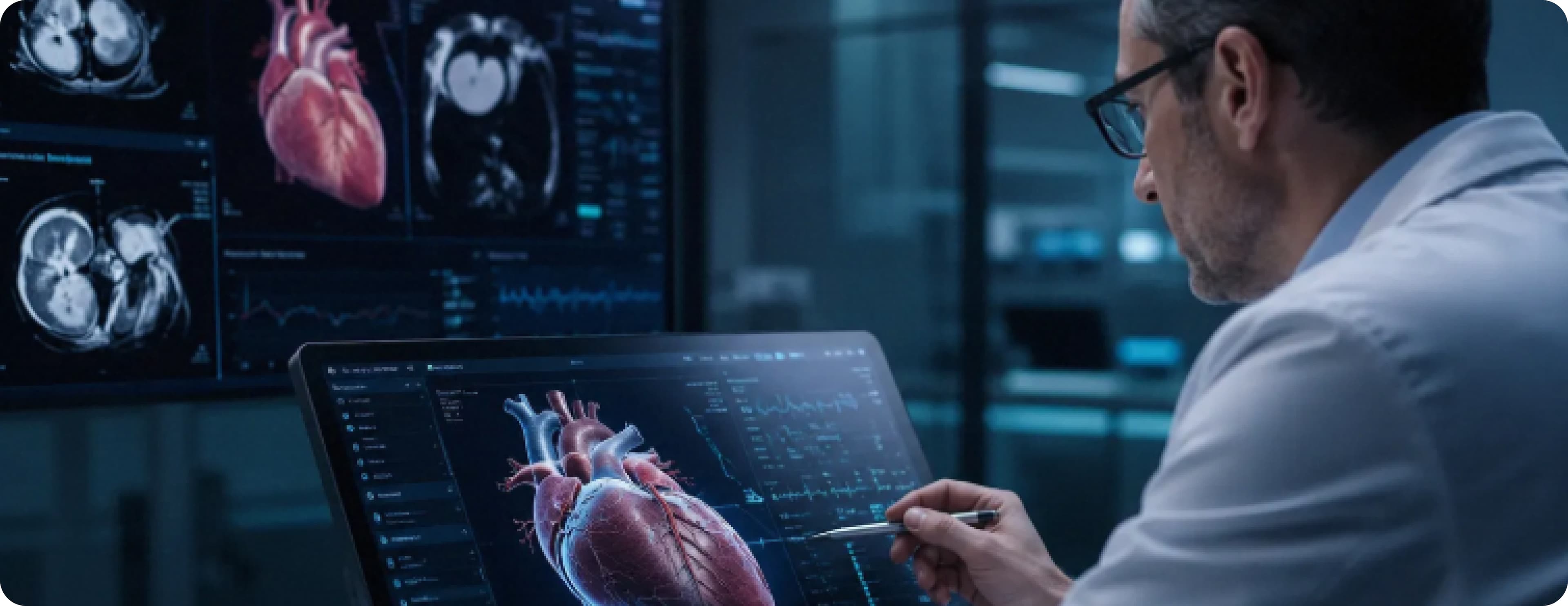
A cardiologist reviewing her morning reading queue has forty-two echocardiograms, seventeen ECGs, and eight Holter monitor reports to interpret before her afternoon clinic begins. Each echocardiogram requires measurement of left ventricular ejection fraction, wall motion assessment, valve function evaluation, and clinical correlation with the referring indication. Each ECG requires rhythm analysis, interval measurement, and pattern recognition across twelve leads. Each Holter report requires arrhythmia burden quantification across twenty-four to forty-eight hours of continuous recording.
She is a skilled cardiologist. She trained for eleven years to perform this work accurately. But the volume she faces every morning is a human cognitive challenge not because any individual study is beyond her expertise, but because the cumulative attention demand of forty-two echocardiograms and seventeen ECGs requires sustained focus that human cognition cannot maintain indefinitely without performance degradation.
At study thirty-one, her attention is not what it was at study one. The subtle wall motion abnormality in the lateral wall that she might have caught with fresh eyes becomes the finding she notes as borderline and moves past. The ECG with the subtle Brugada pattern that requires clinical correlation with the patient's syncope history gets read as normal sinus rhythm with incomplete right bundle branch block.
AI cardiology software addresses this not by replacing her clinical judgment but by being consistently attentive at study forty-two in exactly the way it was at study one, flagging the lateral wall motion abnormality, identifying the Brugada pattern, measuring the QTc interval with precision so that her clinical judgment is applied to findings that AI has already surfaced, rather than to the cognitive work of finding them in the first place.
In 2026, AI cardiology platforms are transforming cardiac imaging interpretation, ECG analysis, cardiac monitoring, heart failure management, and cardiology practice operations across academic medical centers, community hospitals, cardiology group practices, and cardiac telehealth programs.
Key Takeaways
AI cardiology software uses deep learning, computer vision, and predictive analytics to analyze ECGs, echocardiograms, cardiac imaging studies, and continuous monitoring data, providing cardiologists with AI-assisted interpretation support that improves diagnostic accuracy, reduces interpretation time, and ensures consistent pattern detection regardless of reading volume or time of day
Cardiovascular disease remains the leading cause of death in the United States. AI cardiology tools that improve early detection of atrial fibrillation, reduced ejection fraction heart failure, valvular disease, and other cardiovascular conditions directly reduce cardiovascular mortality through earlier diagnosis and treatment
The highest-value AI capabilities are AI ECG interpretation and arrhythmia detection, echocardiogram automated measurement and analysis, cardiac MRI and CT quantification, continuous cardiac monitoring alert management, heart failure risk stratification, and cardiology practice workflow optimization
FDA regulatory classification is central to AI cardiology software development. AI tools that analyze cardiac imaging or ECG data to detect or diagnose specific conditions are regulated as Software as a Medical Device, and most clinically meaningful AI cardiology capabilities require FDA clearance or De Novo authorization before clinical deployment
HIPAA compliance is required throughout cardiac imaging studies; ECG recordings, Holter monitor data, and clinical cardiology records are protected health information requiring full technical safeguards, including DICOM encryption, access controls, and comprehensive audit logging
Integration with PACS, ECG management systems, cardiac monitoring platforms, EHR systems, and cardiac catheterization laboratory information systems creates the connected cardiology ecosystem that makes AI interpretation support a seamless component of cardiologist workflow rather than a separate analytical tool
Total development cost ranges from $80,000 for a focused ECG analysis MVP to $550,000 or more for a full AI-powered cardiology imaging and workflow management platform
What Is AI Cardiology Software?
AI cardiology software is a technology platform that uses artificial intelligence, deep learning, convolutional neural networks, recurrent neural networks, and predictive analytics to support the clinical interpretation, monitoring, and management functions of cardiology practice, providing cardiologists and cardiac care teams with AI-generated analysis of cardiac imaging, ECG recordings, and monitoring data that improves diagnostic accuracy, reduces interpretation time, and identifies clinical findings that require physician attention.
Cardiology is among the medical specialties where AI has demonstrated the clearest clinical value because cardiovascular data (ECGs, echocardiograms, cardiac MRI, CT angiography, Holter monitors) is highly structured, pattern-based, and amenable to the deep learning analysis that excels at finding patterns in high-dimensional data. AI models trained on large datasets of expert-interpreted cardiac studies consistently demonstrate performance approaching or exceeding that of individual cardiologists in specific domains, not because AI understands cardiovascular physiology better than cardiologists, but because AI applies pattern recognition consistently and without the cognitive fatigue that affects human performance at volume.
The clinical impact of AI cardiology software extends beyond interpretation efficiency. AI ECG analysis can detect conditions that are frequently missed in routine clinical ECG interpretation: low ejection fraction heart failure detectable from ECG waveform patterns, early atrial fibrillation in patients with predominantly sinus rhythm, and subtle conduction system disease enabling earlier diagnosis and treatment of conditions where earlier intervention significantly affects outcomes.
Why Is AI Transforming Cardiology in 2026?
What Clinical Problem Does AI Address in Cardiac Interpretation?
Cardiovascular disease is the leading cause of mortality in the United States, responsible for approximately 697,000 deaths annually. Many of the conditions that contribute to cardiovascular mortality are detectable through cardiac testing before they produce life-threatening events: atrial fibrillation that causes thromboembolic stroke, reduced ejection fraction heart failure that causes progressive functional decline, and valvular disease that causes sudden cardiac death. The challenge is detecting these conditions early enough for intervention to change outcomes.
AI cardiology tools that improve the sensitivity of routine cardiac testing for conditions with strong intervention evidence detecting low ejection fraction from standard 12-lead ECG, identifying paroxysmal atrial fibrillation from ambulatory monitoring data, and detecting subclinical hypertrophic cardiomyopathy from echocardiographic patterns create earlier diagnosis opportunities for conditions where earlier treatment demonstrably improves outcomes.
What Interpretation Volume Challenge Makes AI Essential?
The volume of cardiac testing performed in the United States creates an interpretation demand that the cardiologist workforce cannot meet without performance degradation. Approximately 300 million ECGs are performed annually in the US. Approximately 10 million echocardiograms are performed annually. Cardiac monitoring programs for atrial fibrillation detection generate continuous data streams from millions of patients simultaneously. The human cardiologist workforce cannot interpret this volume at the quality level that each individual patient deserves.
AI interpretation support that pre-processes cardiac studies measuring left ventricular ejection fraction from echocardiograms, generating arrhythmia burden summaries from Holter monitors, and identifying findings warranting urgent cardiologist attention from large ECG batches allows cardiologists to apply their clinical expertise to the findings AI has identified rather than to the cognitive work of finding them across high-volume reading queues.
What FDA Regulatory Context Shapes AI Cardiology Development?
The FDA has been active in the AI cardiology regulatory space, clearing multiple AI ECG analysis tools, AI echocardiogram analysis tools, and AI cardiac imaging quantification tools through the 510(k) and De Novo pathways. The regulatory pathway for AI cardiology is well-established, and developers entering this space can learn from the predicate device landscape that existing clearances have created.
For AI cardiology software that makes diagnostic claims AI that detects atrial fibrillation from ECG data, AI that measures left ventricular ejection fraction from echocardiogram video FDA clearance is required before clinical deployment. This regulatory requirement shapes development timelines and investment requirements significantly, and must be addressed before development begins rather than discovered after an AI cardiology product is built.
What Are the Key Use Cases for AI Cardiology Software?
AI ECG Interpretation and Arrhythmia Detection
AI ECG interpretation analyzing 12-lead ECG recordings to identify rhythm abnormalities, conduction abnormalities, ischemic changes, and pattern-based disease detection is the most mature and most widely clinically deployed AI cardiology application. Multiple FDA-cleared AI ECG analysis tools are in clinical use, demonstrating that this AI application has achieved regulatory validation and clinical acceptance.
AI ECG capabilities range from standard arrhythmia detection (atrial fibrillation, ventricular tachycardia, complete heart block, Wolff-Parkinson-White pattern) to novel pattern-based disease detection that goes beyond what traditional ECG interpretation includes. Published research has demonstrated that AI ECG analysis can detect reduced left ventricular ejection fraction from ECG waveform patterns with clinically meaningful sensitivity, identify early hypertrophic cardiomyopathy, and predict atrial fibrillation in patients who are currently in sinus rhythm capabilities that significantly expand the clinical utility of routine ECG recording.
For high-volume ECG programs in primary care clinics, emergency departments, pre-operative assessment programs, and population health screening programs, AI ECG analysis that processes large ECG batches and identifies the studies requiring urgent cardiologist review provides the triage efficiency that makes expert cardiologist review of high-priority findings achievable within the time constraints that clinical urgency requires.
Our AI and ML solutions team builds ECG analysis models with the convolutional neural network architectures, diverse training datasets, and clinical validation standards that FDA-clearable AI ECG tools require.
Echocardiogram Automated Measurement and Analysis
Echocardiogram interpretation: The cardiac ultrasound study that provides the most comprehensive assessment of cardiac structure and function available through non-invasive testing requires measurement of dozens of quantitative parameters (left ventricular ejection fraction, wall thickness, chamber dimensions, diastolic function indices, valvular gradients) alongside qualitative assessment of wall motion, valve morphology, and pericardial pathology.
AI echocardiogram analysis automates the quantitative measurement component of echocardiogram interpretation by measuring left ventricular volumes and ejection fraction from 2D or 3D echo images, quantifying diastolic function parameters, assessing wall motion through segmental motion analysis, and measuring valve orifice areas and gradients. This measurement automation reduces the time cardiologists spend on quantitative measurement and improves measurement consistency by eliminating inter-reader variability in measurement methodology.
For strain imaging analysis, the quantification of myocardial deformation patterns provides sensitive early detection of subclinical cardiomyopathy. AI strain analysis that automatically generates global longitudinal strain measurements from standard echocardiogram acquisitions makes this sensitive functional assessment available without the additional acquisition time and specialized training that manual strain analysis currently requires.
Cardiac MRI Quantification
Cardiac MRI is the gold standard for myocardial characterization, providing tissue characterization capabilities (late gadolinium enhancement for fibrosis detection, T1 and T2 mapping for infiltrative disease, myocardial edema assessment) alongside highly accurate ventricular volume and function quantification. Cardiac MRI analysis is time-consuming; manual segmentation of ventricular contours across multiple phases of the cardiac cycle requires significant post-processing time that limits cardiac MRI throughput.
AI cardiac MRI quantification automates ventricular segmentation for volume and ejection fraction calculation, automated late gadolinium enhancement detection and quantification, and tissue characterization pattern classification, reducing cardiac MRI post-processing time and providing the quantitative outputs that clinical reporting requires without the manual segmentation burden that currently limits cardiac MRI utilization.
Cardiac CT Analysis
Cardiac CT, including coronary CT angiography for coronary artery disease assessment and CT calcium scoring for cardiovascular risk stratification, generates image data that AI analysis can quantify more consistently and more rapidly than manual review. AI coronary CT angiography analysis quantifying stenosis severity, plaque burden, and plaque characterization across the coronary tree provides the systematic quantitative reporting that manual CT angiography interpretation cannot consistently deliver.
For CT calcium scoring, specifically a high-volume population health screening application where AI quantification of coronary artery calcium provides cardiovascular risk stratification, AI scoring that provides accurate, reproducible Agatston scores from non-contrast cardiac CT images makes calcium scoring practical for primary care and population health screening programs at volume levels that manual scoring cannot support.
Continuous Cardiac Monitoring and Alert Management
Continuous cardiac monitoring, ambulatory ECG monitoring for atrial fibrillation detection, implantable loop recorder data analysis, and wearable cardiac monitoring data streams generate enormous volumes of cardiac rhythm data that require intelligent triage to surface the findings requiring physician attention from the background of normal rhythm.
AI cardiac monitoring analysis processes continuous rhythm data streams to identify arrhythmia events, quantify arrhythmia burden, classify arrhythmia types, and generate prioritized alert queues for cardiologist review, providing the analytical infrastructure that makes continuous monitoring programs clinically manageable. Without AI triage, the volume of monitoring data generated by large cardiac monitoring programs exceeds the physician review capacity available for comprehensive manual interpretation.
Our EHR and EMR integration practice builds the clinical data integrations that connect AI cardiology analysis outputs to the cardiologist's clinical workflow, making AI-identified findings accessible within the EHR rather than requiring cardiologists to access separate analytical platforms.
Heart Failure Risk Stratification and Management
Heart failure affects approximately 6.2 million Americans and is responsible for approximately one million hospitalizations annually, a condition where proactive risk stratification and between-visit monitoring can significantly reduce hospitalization events that most affect patient quality of life and healthcare resource utilization.
AI heart failure risk stratification analyzes clinical data, including echocardiographic parameters, laboratory values, medication adherence, remote monitoring device data, and prior hospitalization history, to generate patient-specific hospitalization risk scores that enable care teams to intensify management for high-risk patients before decompensation occurs. AI-identified high-risk patients receive proactive outreach, medication optimization review, and monitoring intensification that reduces hospitalization rates in ways that fixed-protocol management cannot achieve.
What Are the Key Features of AI Cardiology Software?
AI ECG Analysis Engine
Deep learning ECG interpretation with rhythm classification, conduction abnormality detection, ischemia pattern identification, QTc measurement, and novel pattern-based disease detection. FDA-clearable analysis architecture with predicate device identification and clinical validation study design appropriate for 510(k) submission.
Echocardiogram AI Analysis
Automated left ventricular volume and ejection fraction measurement, wall motion segmental analysis, diastolic function parameter quantification, valve gradient measurement, and global longitudinal strain analysis. DICOM integration for echo image acquisition and report generation with AI-populated quantitative measurements.
Cardiac MRI and CT Quantification
Automated ventricular segmentation for cardiac MRI quantification, late gadolinium enhancement detection and quantification, CT calcium scoring, and coronary CT angiography stenosis quantification. Integration with cardiac imaging PACS for automated study processing and AI-populated quantitative reports.
Continuous Monitoring Alert Management
Cardiac rhythm data stream processing for ambulatory ECG monitoring, implantable device data analysis, and wearable monitoring data triage. AI-generated prioritized alert queues with arrhythmia type classification and burden quantification for cardiologist review workflow management.
DICOM and PACS Integration
Bidirectional DICOM integration for cardiac imaging study receipt, AI analysis processing, and AI-populated report generation. PACS integration for study routing to AI processing and AI result delivery to cardiologist reading workstations.
Cardiology Practice Workflow Management
Study reading queue management with AI-generated priority scoring, reading assignment optimization for subspecialty expertise matching, turnaround time monitoring, and cardiologist productivity analytics.
Heart Failure Risk Dashboard
Population-level heart failure risk stratification with AI-generated risk scores, high-risk patient alert generation, remote monitoring data integration, and care management workflow tools for heart failure program coordinators.
HIPAA-Compliant Data Architecture
All cardiology data ECG recordings, echocardiogram studies, cardiac MRI and CT images, monitoring data, and clinical records is protected health information requiring AES-256 encryption, role-based access controls, DICOM TLS for cardiac imaging transmission, and BAAs with all integrated systems.
Our HIPAA-compliant software development practice builds compliance architecture for cardiology AI platforms handling cardiac imaging studies, ECG recordings, and comprehensive cardiac clinical records.
Cardiology Analytics Dashboard
Reading queue performance analytics, AI analysis accuracy monitoring, turnaround time reporting, arrhythmia detection rate trends, and heart failure program outcome metrics provide cardiology department directors and practice administrators with the operational intelligence for systematic performance management.
Our healthcare UI/UX design team designs cardiology analytics dashboards tested with real cardiologists, cardiac imaging directors, and cardiology practice administrators.
How to Build AI Cardiology Software: Step by Step
Step 1: Define the Clinical Scope and FDA Pathway
Building AI cardiology software begins with defining precisely which cardiac analysis functions the AI will perform and whether each function constitutes a diagnostic claim requiring FDA clearance.
AI that measures left ventricular ejection fraction from echocardiogram video and presents the measurement as a clinical parameter requires FDA clearance as a measurement device. AI that detects atrial fibrillation from ECG data and presents the finding for clinical action requires FDA clearance as a diagnostic decision support tool. Regulatory counsel with FDA digital health experience must assess each planned AI function before development begins because the FDA regulatory pathway determines development requirements, validation study design, and time-to-market timelines.
Step 2: Secure Cardiac Training Data
AI cardiac analysis model performance is directly determined by the quality, volume, and diversity of training data. ECG analysis models require large datasets of ECG recordings with expert cardiologist interpretation labels across the full range of ECG findings the model is trained to detect. Echocardiogram analysis models require expert-labeled echo video datasets with measurements verified by experienced cardiac imagers.
Training data diversity across patient demographics, recording equipment types, image acquisition protocols, and clinical settings determines whether the model generalizes to the full range of patients and equipment it will encounter in clinical deployment. Data that overrepresents specific patient populations or equipment types produces models with systematic performance gaps for underrepresented groups.
Step 3: Address FDA Regulatory Requirements
For AI cardiology features requiring FDA clearance, design the development process to generate the clinical validation evidence FDA review requires: analytical validation studies demonstrating model performance on labeled test datasets, and, where required by the specific device type, clinical validation studies demonstrating the AI tool's performance in clinical practice settings.
Our MVP and product strategy process addresses FDA regulatory pathway planning, predicate device identification, and clinical validation study design as core discovery phase components for AI cardiology development.
Step 4: Build DICOM and Cardiac Data Integration
Build the DICOM integration infrastructure: DICOM C-STORE for cardiac imaging study receipt, DICOM Structured Report for AI result delivery, DICOM TLS for encrypted cardiac imaging transmission, and integration with cardiac-specific PACS platforms. Build ECG data integration using SCP-ECG and MUSE XML formats for ECG system connectivity.
Step 5: Develop and Validate AI Models
Develop AI analysis models using the deep learning architectures most appropriate for each cardiac data type. Train models on curated, labeled cardiac datasets. Validate model performance on independent held-out test sets with demographic subgroup analysis. Conduct the analytical validation studies required for FDA submission for regulated device functions.
Step 6: Build Clinical Workflow Integration
Build the clinical workflow integration: AI analysis results delivery to cardiologist reading workstations, AI-populated report templates for cardiologist review and attestation, reading queue management with AI priority scoring, and EHR integration for AI result documentation in the clinical record.
Step 7: Build Monitoring and Alert Management
For continuous monitoring applications, build the real-time cardiac data stream processing infrastructure for arrhythmia event detection, burden quantification, alert classification, and cardiologist alert queue management with priority-based worklist organization.
Step 8: Implement HIPAA Compliance Architecture
Build the full HIPAA compliance architecture, with encrypted storage and transmission for all cardiac imaging and ECG data, including DICOM TLS, role-based access controls reflecting cardiology department roles, comprehensive audit logging, and BAAs with all integrated PACS, ECG management, and EHR systems.
Step 9: Pilot and Measure
Deploy in a structured clinical pilot measuring AI analysis accuracy against cardiologist reference interpretations, cardiologist reading time per study with AI assistance, critical finding detection rate, and cardiologist satisfaction with AI workflow integration.
Our DevOps and cloud solutions team builds the deployment infrastructure, AI model performance monitoring, DICOM pipeline management, and cardiology analytics that keep the platform accurate and FDA-compliant as patient populations and cardiac imaging technology evolve.
What Technology Powers AI Cardiology Software?
Deep Learning for Cardiac Analysis
PyTorch is the primary deep learning framework for AI cardiology model development. For ECG analysis, detecting arrhythmias and cardiac patterns from 12-lead ECG recordings, 1D convolutional neural networks and transformer-based sequence models trained on large ECG datasets with expert interpretation labels produce state-of-the-art arrhythmia detection and pattern classification performance.
For echocardiogram analysis measuring ventricular volumes, ejection fraction, and wall motion, 2D and 3D echo video spatiotemporal convolutional networks and video transformers process the temporal sequence of cardiac motion that echocardiogram video captures. EchoNet-Dynamic and similar architectures pre-trained on large echocardiogram datasets provide the transfer learning foundation that reduces labeled training data requirements for specific measurement tasks.
For cardiac MRI analysis, ventricular segmentation across cardiac phases for volume quantification: U-Net-based segmentation architectures with multi-scale feature extraction provide accurate myocardial boundary delineation. For CT calcium scoring, detecting and quantifying coronary calcium from non-contrast cardiac CT, 3D convolutional networks trained on CT calcium scoring datasets provide automated Agatston score calculation.
Cardiac Data Integration Infrastructure
DICOM for all cardiac imaging data: DICOM C-STORE and C-FIND for study management, DICOM Structured Report for AI result delivery, and DICOM TLS for encrypted transmission. SCP-ECG (Standard Communications Protocol for Computer-Assisted Electrocardiography) and MUSE XML for ECG system integration. HL7 FHIR Observation and DiagnosticReport resources for AI result documentation in EHR clinical records.
Backend Infrastructure
Python with FastAPI for the primary API layer. PostgreSQL for structured cardiology workflow and clinical data. AWS S3 with server-side encryption for cardiac imaging study storage. Redis for real-time monitoring alert queue management and reading worklist state. Apache Kafka for high-volume continuous monitoring data stream processing.
Cloud Infrastructure
AWS with a HIPAA Business Associate Agreement. Amazon S3 with server-side encryption for DICOM cardiac imaging storage. Amazon SageMaker for AI model training and serving. Amazon HealthLake for FHIR-based clinical data management. AWS CloudTrail for comprehensive HIPAA audit logging.
What Are the HIPAA and FDA Compliance Requirements?
What HIPAA Requirements Apply to Cardiology AI?
Cardiac imaging studies, ECG recordings, Holter monitor data, and clinical cardiology records are all protected health information requiring full HIPAA technical safeguards. DICOM cardiac imaging transmission requires DICOM TLS rather than unencrypted DICOM transport. Cardiac imaging storage in cloud environments requires HIPAA-eligible storage services with signed BAAs. Role-based access controls must reflect cardiology department role structures: cardiologists access reading queues and full clinical data, technologists access acquisition workflow, and billing staff access procedure codes without clinical imaging access.
What FDA Requirements Apply to AI Cardiology Software?
The FDA regulates AI cardiology tools that detect, diagnose, or measure specific cardiac conditions or parameters as Software as a Medical Device. AI ECG analysis that detects atrial fibrillation for clinical action requires FDA clearance. AI echocardiogram analysis that measures left ventricular ejection fraction as a clinical parameter requires FDA clearance. AI cardiac CT analysis that quantifies coronary stenosis severity requires FDA clearance.
The 510(k) pathway demonstrating substantial equivalence to a predicate cleared device is the most common regulatory pathway for AI cardiology tools with existing predicate devices. The De Novo pathway applies to novel AI cardiology tools without appropriate predicates. FDA Software as a Medical Device Pre-Submission meetings allow developers to discuss regulatory strategy with FDA before formal submission, and this interaction is strongly recommended for AI cardiology development programs to confirm regulatory pathway and validation study requirements before significant development investment.
What Are the Common Mistakes to Avoid?
1. FDA Classification After Development
The most expensive mistake in AI cardiology development is building AI analysis features that make diagnostic claims atrial fibrillation detection, ejection fraction measurement without obtaining FDA regulatory pathway assessment before development begins. Features that require FDA clearance must be developed with the clinical validation documentation that FDA review requires, and this documentation must be planned and executed prospectively during development rather than constructed retroactively after a product is built.
2. Training Data Without Demographic Diversity Validation
AI cardiac analysis models trained on datasets that underrepresent specific demographic groups elderly patients, women, patients with specific comorbidities, patients recorded on lower-quality equipment produce systematic performance gaps for underrepresented populations. Cardiac AI that performs well for the majority patient population but misses atrial fibrillation or reduced ejection fraction in specific demographic groups creates clinical risk rather than improving it. Training data diversity assessment and demographic subgroup performance validation are non-negotiable quality requirements.
3. AI Results Without Cardiologist Attestation Workflow
AI cardiology analysis systems that generate results and document them in the clinical record without cardiologist review and attestation create both clinical and regulatory risk; the AI result is being used as a clinical finding without the physician oversight that regulated medical device use requires. AI cardiology results must be presented for cardiologist review and require explicit cardiologist attestation before documentation as clinical findings.
4. DICOM Integration Without Conformance Testing
DICOM integration that has not been tested against the specific DICOM conformance statements of the cardiac imaging modalities and PACS systems in the target environment produces integration failures in clinical deployment. DICOM conformance testing against each target system's conformance statement is required before clinical deployment.
5. No Model Performance Monitoring Post-Deployment
AI cardiology model performance can degrade over time as imaging technology, patient population characteristics, and clinical protocols evolve. AI models deployed without ongoing performance monitoring comparing AI output against cardiologist reference interpretations on a continuous sampling basis accumulate performance drift that is not detected until clinical quality review identifies AI-specific errors.
How Does Codieshub Build AI Cardiology Software?
At Codieshub, we build AI cardiology software for cardiology practices, hospital cardiology departments, cardiac imaging centers, and health tech companies that need cardiology AI platforms designed for the specific FDA regulatory requirements, DICOM integration complexity, clinical validation standards, and cardiologist workflow integration demands of cardiac AI development.
Every engagement begins with our MVP and product strategy process, which addresses clinical scope definition, FDA regulatory pathway assessment with predicate device identification, cardiac training data strategy, DICOM integration architecture, clinical validation study design, and HIPAA compliance design before production code is written.
Our AI and ML solutions team builds ECG analysis models, echocardiogram quantification systems, cardiac MRI and CT analysis tools, and continuous monitoring analytics with demographic diversity validation, FDA-appropriate analytical validation documentation, and model performance monitoring built in from the beginning.
Our API integration services team builds DICOM integrations with cardiac PACS platforms and ECG system integrations using SCP-ECG and MUSE XML. Our EHR and EMR integration team builds HL7 FHIR integrations for AI cardiology result documentation in EHR clinical records. Our healthcare UI/UX design team designs cardiologist reading workflow interfaces and cardiology analytics dashboards tested with real cardiologists and cardiac imaging directors. Our HIPAA-compliant software development practice ensures full compliance, including DICOM TLS for cardiac imaging transmission. And our DevOps and cloud solutions team builds the deployment infrastructure, AI model serving at clinical imaging scale, DICOM pipeline management, and performance monitoring that keep the cardiology AI platform accurate and FDA-compliant as cardiac imaging technology and patient populations evolve.
Conclusion
Cardiovascular disease is the leading cause of death in the United States, and the most powerful tools available for reducing cardiovascular mortality earlier detection of atrial fibrillation, earlier identification of reduced ejection fraction heart failure, and more sensitive screening for valvular disease and cardiomyopathy are already present in the cardiac testing that the healthcare system performs at enormous scale every year. The gap is not in the tests being performed. The gap is in the consistency and completeness with which the findings that tests contain are identified, surfaced, and acted upon.
AI cardiology software closes this gap not by replacing the cardiologist whose clinical judgment determines what findings mean for each patient's care, but by ensuring that the pattern recognition that identifies those findings is applied consistently and completely across every study in the reading queue. The cardiologist reviewing study forty-two of forty-two deserves the same quality of AI-assisted pattern detection that study one received, and so does the patient whose diagnosis depends on it.
At Codieshub, we build AI cardiology software for cardiology practices, cardiac imaging programs, and health tech companies that understand what cardiac AI requires: FDA regulatory precision, deep learning models trained on diverse cardiac datasets with demographic equity validation, DICOM integration rigor, and cardiologist workflow integration that makes AI assistance a seamless component of clinical practice rather than an additional system to manage.
Ready to build AI cardiology software that improves diagnostic accuracy and practice efficiency? Schedule a Discovery Call to tell us about your cardiology AI scope and clinical priorities, and we will send you a tailored development and regulatory game plan within 48 hours.
Frequently Asked Questions
1. What is AI cardiology software?
AI cardiology software uses deep learning and computer vision to analyze ECG recordings for arrhythmia detection and pattern-based disease identification, measure echocardiogram parameters including ejection fraction and wall motion, quantify cardiac MRI and CT findings, process continuous monitoring data streams for arrhythmia alert management, stratify heart failure risk, and manage cardiology practice workflow, providing cardiologists with consistent, high-quality AI analysis support across high-volume cardiac interpretation workloads.
2. How does AI ECG analysis improve cardiology practice efficiency?
AI ECG analysis processes large ECG batches and identifies studies containing findings requiring urgent cardiologist attention, separating normal or routine studies from those with critical arrhythmias, ischemic patterns, or pattern-based disease indicators. Cardiologists apply their clinical expertise to the AI-identified findings rather than to the cognitive work of finding them across high-volume reading queues, improving both interpretation efficiency and the consistency of critical finding detection regardless of reading queue position or time of day.
3. Does AI cardiology software require FDA clearance?
Yes, for most clinically meaningful AI cardiology capabilities. AI tools that detect or diagnose specific cardiac conditions atrial fibrillation detection from ECG, reduced ejection fraction identification from echocardiogram, or measuring specific clinical parameters require FDA clearance as Software as a Medical Device. The 510(k) pathway demonstrating substantial equivalence to a predicate cleared device is the most common pathway. FDA regulatory counsel assessment before development begins is essential; building regulated features without FDA pathway planning creates post-development regulatory requirements that are significantly more expensive to address.
4. How does AI echocardiogram analysis improve cardiac imaging workflows?
AI echocardiogram analysis automates the quantitative measurement component of echo interpretation, measuring left ventricular volumes and ejection fraction, quantifying diastolic function parameters, assessing segmental wall motion, and calculating valve parameters from echo images. This automation reduces per-study cardiologist time on quantitative measurement, improves measurement consistency by eliminating inter-reader variability, and provides global longitudinal strain measurements from standard echo acquisitions without additional acquisition time.
5. How does DICOM integration work in AI cardiology software?
AI cardiology software receives cardiac imaging studies through DICOM C-STORE from PACS or directly from imaging modalities, processes studies through AI analysis models, and delivers AI-generated measurements and findings through DICOM Structured Reports to cardiologist reading workstations. DICOM TLS provides encrypted DICOM transmission for HIPAA-compliant cardiac imaging transfer. DICOM conformance testing against each target PACS and modality system's conformance statement is required before clinical deployment to ensure reliable integration across the diversity of DICOM implementations in clinical cardiology environments.
6. How does AI continuous monitoring management support cardiac arrhythmia programs?
AI cardiac monitoring analysis processes continuous rhythm data streams from ambulatory ECG monitors, implantable loop recorders, and wearable devices, detecting arrhythmia events, classifying arrhythmia types, quantifying arrhythmia burden, and generating prioritized cardiologist alert queues organized by clinical urgency. This triage infrastructure makes large-scale cardiac monitoring programs clinically manageable by focusing cardiologist review on the AI-identified events requiring attention rather than requiring manual review of continuous data streams from hundreds of simultaneous monitoring patients.
7. How long does it take to build AI cardiology software?
A focused MVP with AI ECG arrhythmia detection and cardiologist review workflow takes four to eight months. A mid-level platform with ECG analysis, echocardiogram quantification, monitoring management, and EHR integration takes eight to sixteen months. A full enterprise AI cardiology platform with comprehensive cardiac imaging AI, FDA clearance preparation across all regulated functions, and multi-site deployment takes eighteen to thirty-six months. FDA regulatory pathway preparation and clinical validation study execution are the primary additional timeline drivers beyond standard software development.
8. How much does AI cardiology software cost to build?
A focused MVP costs $80,000 to $160,000. A mid-level AI cardiology platform costs $160,000 to $340,000. A full enterprise AI cardiology imaging and management platform costs $340,000 to $550,000 or more. Annual maintenance typically costs $50,000 to $120,000. Primary cost drivers are deep learning model development on diverse cardiac training datasets, FDA regulatory preparation and clinical validation study execution, DICOM integration and conformance testing across cardiac imaging systems, demographic diversity validation for equitable model performance, and cardiologist attestation workflow design meeting FDA intended-use requirements.